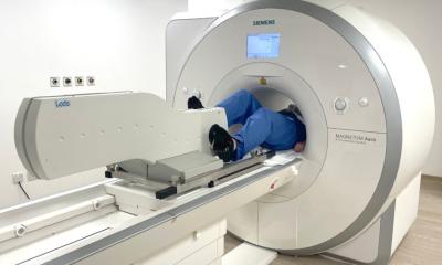
If we make them exercise during their MRI they can no longer compensate and we get a much clearer idea of who has the least resilience and who is really at risk.
— Declan O’Regan
Investigating the heart’s response to exercise has taken a significant leap forward through the development of advanced imaging technologies. One such groundbreaking approach is ExCMR, which allows healthcare professionals to obtain live cardiac imaging while patients engage in physical activity. This dynamic method provides a fresh perspective on cardiovascular health, particularly as it varies between different demographics, such as men and women.
Recently, a team of researchers embarked on a unique study involving 161 healthy participants aged between 22 and 77. Their goal? To observe how a healthy heart reacts during exercise. This investigation revealed crucial differences in heart responses between genders. Notably, men exhibited a more pronounced cardiovascular response to exercise than women, even when accounting for variables such as body size. This insight is vital; it underscores the necessity to tailor heart health diagnostics and treatments based on gender-specific responses.
ExCMR stands for Exercise Cardiac Magnetic Resonance imaging. This innovative method entails a specialized attachment that enables participants to pedal—much like riding a stationary bike—while lying in the MRI scanner. As individuals pedal, their hearts’ responses are closely monitored, allowing for real-time assessment of heart health. This technique is particularly advantageous because, traditionally, MRI scans are performed when individuals are at rest, which may not reflect their true cardiovascular condition.
The reality of heart disease is that many individuals, particularly younger people or those in the early stages of the illness, may not exhibit obvious symptoms during rest. Often, the underlying condition becomes more pronounced during everyday activities such as climbing stairs or rushing for a bus. According to Professor Declan O’Regan, head of the computational cardiac imaging group at the LMS, many patients are adept at compensating for their cardiovascular issues while at rest. However, when subjected to exercise during an MRI, these compensatory mechanisms are disrupted. This opens up a window of opportunity to assess their true cardiac resilience, ultimately identifying those who are genuinely at risk.
This novel approach to cardiac imaging holds promise for earlier and more accurate diagnoses of heart conditions. By capturing the heart’s performance in a state of exertion, healthcare providers can better distinguish between a healthy heart and one that may be operating under stress due to an underlying problem. Such advancements not only aim to enhance diagnostic accuracy but also have the potential to improve treatment outcomes for patients experiencing heart disease.
In summary, the integration of exercise into cardiac imaging technologies like ExCMR signifies a paradigm shift in assessing cardiovascular health. By focusing on the heart’s response during physical activity, researchers and clinicians are paving the way for more nuanced and effective approaches to diagnosing and managing heart conditions. With this emerging understanding, the future of cardiovascular care looks to be not only more informed but also more proactive.


